Experiencing pain in the inside of knee can turn simple daily tasks—like walking down stairs, getting out of a chair, or enjoying a brisk stroll—into a painful struggle. Known clinically as medial knee pain, discomfort along the inner aspect of the joint is one of the most common musculoskeletal complaints treated by orthopedic specialists and physical therapists worldwide.
Because the knee is a complex hinge joint bearing up to four times your body weight with every step, even minor irritation to its inner structural components can result in persistent discomfort, stiffness, or functional instability. Understanding the root cause of your pain in the inside of knee is the vital first step toward restoring mobility and achieving lasting, long-term joint comfort.
If you are looking for an effective, holistic approach to rebuild joint integrity and reduce systemic inflammation alongside standard conservative care, incorporating a proven natural joint support formula into your routine can accelerate recovery and protect cartilage health.
Anatomy of the Medial Knee: What Lies Beneath?
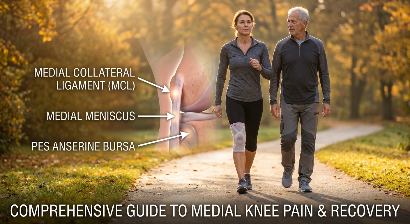
To understand why pain in the inside of knee occurs, it helps to examine the delicate anatomy of the medial compartment. The knee connects the thigh bone (femur) to the shin bone (tibia), anchored and cushioned by several specialized tissues:
-
Medial Collateral Ligament (MCL): A broad, thick band of fibrous tissue running along the inner side of the knee joint. Its primary function is to prevent the knee from collapsing inward toward the opposite leg (valgus stress).
-
Medial Meniscus: A C-shaped piece of tough, rubbery fibrocartilage that sits between the femur and tibia on the inner side. It acts as a primary shock absorber, distributing weight evenly across the joint surface.
-
Pes Anserine Tendons and Bursa: Located roughly two inches below the inner knee joint line, this area is where three key tendons (sartorius, gracilis, and semitendinosus) insert into the tibia. A small fluid-filled sac called a bursa cushions these tendons against friction.
-
Articular Cartilage: A smooth, slippery tissue covering the ends of the femur and tibia within the medial compartment, allowing the bones to glide effortlessly against one another.
When any of these soft tissues or articular structures become strained, inflamed, torn, or degraded, the result is sharp or dull pain in the inside of knee.
Primary Causes of Pain in the Inside of the Knee
Medial knee discomfort rarely appears without a specific cause. It typically falls into two primary categories: acute traumatic injuries or chronic overuse and degenerative conditions.
                        MEDIAL KNEE PAIN
                               |
       +-----------------------+-----------------------+
       |                                               |
 ACUTE INJURIES                               CHRONIC / OVERUSE
 - MCL Sprain/Tear                            - Medial Knee Osteoarthritis
 - Medial Meniscus Tear                       - Pes Anserine Bursitis
 - Traumatic Bone Bruise                      - Medial Plica Syndrome
                                              - Overpronation (Foot Biomechanics)
1. Medial Collateral Ligament (MCL) Sprain or Tear
An MCL injury usually occurs due to a direct blow to the outer side of the knee or a sudden twisting movement while the foot is planted firmly on the ground. Common in sports like soccer, basketball, skiing, and football, an MCL strain causes immediate pain in the inside of knee, local swelling, tenderness along the inner joint line, and a feeling of joint instability when bearing weight.
2. Medial Meniscus Tear
The inner meniscus is anchored more firmly than the outer meniscus, making it considerably more vulnerable to injury. A medial meniscus tear often results from twisting the knee while pivoting or squatting. In older adults, degenerative meniscus tears can occur with minimal force due to age-related tissue weakening. Typical symptoms include a clicking or catching sensation, stiffness, mild swelling, and persistent pain in the inside of knee during deep bending or twisting.
3. Pes Anserine Bursitis
Pes anserine bursitis refers to inflammation of the bursa located between the tibia and the three inserting inner thigh tendons. This condition is particularly common among runners, cyclists, individuals with tight hamstrings, and those living with knee osteoarthritis. It manifests as a localized, tender ache roughly two inches below the inner joint line, often worsening when climbing stairs or rising from a seated position.
4. Medial Compartment Knee Osteoarthritis
Because human gait biomechanics naturally transfer a greater portion of weight onto the inner side of the knee joint, medial knee osteoarthritis is the most frequent form of knee arthritis. Over time, the protective articular cartilage wears down, causing bone-on-bone friction, osteophyte (bone spur) formation, morning stiffness lasting under 30 minutes, and a deep, aching pain in the inside of knee that worsens with prolonged standing or walking.
5. Medial Plica Syndrome
Plica are remnants of fetal tissue folds within the synovial capsule of the knee. In some individuals, the medial plica becomes thickened, irritated, or pinched between the kneecap and thigh bone due to repetitive bending or sudden increases in physical activity. This leads to a distinct catching sensation, mild swelling, and localized inner knee pain.
Symptom Comparison Table
Identifying the characteristics of your symptoms can help narrow down the underlying cause of your pain in the inside of knee:
| Condition | Onset Type | Key Characteristics | Common Triggers |
| MCL Sprain | Sudden / Acute | Pop sound, inner knee swelling, instability | Sideways impact, twisting foot |
| Medial Meniscus Tear | Sudden or Gradual | Catching, locking, localized inner joint line tenderness | Pivoting, deep squatting |
| Pes Anserine Bursitis | Gradual / Overuse | Localized tenderness 2 inches below joint line | Stair climbing, running, tight hamstrings |
| Medial Osteoarthritis | Slow / Chronic | Morning stiffness, crepitus, deep ache after activity | Weight-bearing, prolonged walking |
| Medial Plica Syndrome | Gradual | Snapping sensation, aching along inner kneecap border | Repetitive bending, cycling |
Diagnostic Approach: How to Evaluate Your Knee Pain
When evaluating pain in the inside of knee, healthcare providers combine a structured medical history with targeted physical examination techniques:
-
Palpation Test: Pressing along the inner joint line to isolate tenderness at the MCL insertion versus the pes anserine bursa or meniscus.
-
Valgus Stress Test: Pushing the outer knee inward while stabilizing the ankle to test the structural integrity and resistance of the MCL.
-
McMurray Test: Flexing, rotating, and extending the knee joint to elicit clicking or pain indicative of a medial meniscus tear.
-
Diagnostic Imaging:
-
X-rays: Useful for evaluating bone alignment, joint space narrowing, bone spurs, and structural signs of medial knee osteoarthritis.
-
MRI (Magnetic Resonance Imaging): The gold standard for visualizing soft tissues including the MCL, meniscus, cartilage, and bursa.
-
Conservative Treatment Strategies for Inner Knee Relief
Fortunately, the vast majority of non-traumatic causes of pain in the inside of knee respond remarkably well to non-surgical conservative care. Implementing a structured recovery plan allows injured tissues to heal while reducing local mechanical friction.
The Modified R.I.C.E. Protocol
For acute flare-ups or sudden injuries, initiate conservative management immediately:
-
Rest: Avoid activities that provoke sharp discomfort, such as high-impact jumping, sprinting, or heavy squatting. Switch temporarily to low-impact swimming or upper-body workouts.
-
Ice: Apply a cold pack wrapped in a thin towel to the medial knee for 15 to 20 minutes, 3 to 4 times daily, to minimize localized inflammation and numb sharp pain signals.
-
Compression: Use an elastic knee sleeve or supportive bandage around the joint to suppress swelling and enhance proprioception (joint position sense).
-
Elevation: Prop your leg up above heart level on pillows while resting to facilitate venous return and fluid lymphatic drainage.
Exercises and Stretches to Rebuild Knee Stability
Strengthening the muscles surrounding the knee while restoring muscular flexibility plays a pivotal role in relieving long-term pain in the inside of knee.
                           REHABILITATION PROGRAM
                                      |
       +------------------------------+------------------------------+
       |                                                             |
 STRENGTHENING                                                STRETCHING
 - Straight Leg Raises (VMO focus)                            - Adductor Stretch
 - Clamshells (Gluteus Medius)                                - Hamstring Wall Stretch
 - Step-Downs (Eccentric Control)                             - Calf/Soleus Stretch
1. VMO (Vastus Medialis Oblique) Straight Leg Raise
The inner portion of your quadriceps (the VMO) helps pull the kneecap smoothly along its track and supports inner joint stability.
-
Lay flat on your back with one knee bent and the affected leg fully extended.
-
Rotate your foot slightly outward (externally) at a 45-degree angle.
-
Engage your quad and slowly lift your leg 12 inches off the ground.
-
Hold for 3–5 seconds, then lower slowly. Perform 3 sets of 12 repetitions.
2. Gluteus Medius Clamshells
Weak hip abductors cause the thigh to collapse inward during walking, adding excessive stress to the medial knee compartment.
-
Lie on your uninjured side with knees bent at a 90-degree angle and feet stacked.
-
Keeping your heels pressed together, raise your top knee upward as high as possible without rotating your pelvis backward.
-
Lower slowly. Perform 3 sets of 15 repetitions per side.
3. Inner Thigh (Adductor) Gentle Stretch
Tight inner thigh muscles pull on the medial tendon attachments, exacerbating pes anserine bursitis.
-
Sit tall on the floor in a butterfly position with the soles of your feet touching.
-
Hold your feet and gently allow your knees to fall outward toward the floor without forcing them.
-
Hold the comfortable stretch for 30 seconds while breathing deeply. Repeat 3 times.
Nutrition, Inflammation, and Internal Joint Support
While targeted physical rehabilitation addresses structural alignment, internal tissue healing relies heavily on biochemistry. Chronic low-grade inflammation inside the joint fluid can break down collagen structures and accelerate cartilage degradation, keeping your knee stuck in a painful cycle.
To support healthy cartilage synthesis and damp down joint inflammation:
-
Adopt an Anti-Inflammatory Diet: Emphasize wild-caught fatty fish (rich in Omega-3 fatty acids), leafy green vegetables, berries rich in polyphenols, extra virgin olive oil, and turmeric.
-
Optimize Hydration: Articular cartilage is composed of up to 80% water. Consistent cellular hydration keeps joint cartilage resilient to daily compressive forces.
-
Targeted Nutritional Supplementation: Essential micronutrients, bioactive collagen peptides, glucosamine, and botanical extracts can significantly boost your body’s innate ability to repair damaged joint tissue. Utilizing an all-natural, scientifically formulated knee and joint restoration blend provides the necessary building blocks to rebuild worn cartilage and alleviate chronic stiffness naturally.
Biomechanics and Prevention: Protecting Your Inner Knee
To keep pain in the inside of knee from returning once you have recovered, pay close attention to your body mechanics and footwear:
-
Correct Overpronation: When your foot arches collapse inward (overpronation) during gait, it forces your tibia to rotate internally, putting excessive stretch on the MCL and squeezing the medial meniscus. Consider custom or off-the-shelf supportive orthotic insoles.
-
Replace Worn Footwear: Athletic shoes lose up to 50% of their shock-absorbing capabilities after 300 to 500 miles of use. Replacing worn shoes prevents unnecessary force transmission to your knees.
-
Maintain Healthy Body Weight: Every extra pound of body weight exerts roughly four pounds of additional pressure on your knee joints with every single step. Achieving and maintaining a healthy weight significantly decreases compressive strain on the inner knee compartment.
When to Seek Immediate Medical Attention
While mild pain in the inside of knee can often be managed effectively at home, certain “red flag” symptoms warrant immediate evaluation by an orthopedic surgeon or emergency medical provider:
⚠️ Red Flag Symptoms:
Inability to bear any weight on the affected leg.
Visibly deformed joint contour or knee dislocation appearance.
A loud “pop” accompanied by rapid, severe swelling within 1 to 2 hours of injury.
Inability to fully extend or bend the knee (joint locking/mechanically blocked).
High fever, redness, warmth, or severe throbbing, which may indicate a septic joint infection.
Reclaiming Pain-Free Movement
Living with pain in the inside of knee does not have to be a permanent reality. By accurately identifying the underlying cause—whether an acute MCL strain, a medial meniscus tear, pes anserine bursitis, or early medial knee osteoarthritis—you can combine gentle physical therapy, biomechanical corrections, and optimal nutrition to kickstart recovery.
Be patient with your body throughout the restoration process. Pair targeted strengthening exercises with a high-quality advanced joint health formula to reduce joint discomfort, nourish your cartilage, and return to an active, pain-free life.